
"I honestly can’t remember what I had injected back then. All I remember is the doctor saying it would absorb." Other people have had fillers, fat grafting and several corrections on the same face at different clinics, and can no longer tell which area belongs to which treatment. Many start guessing brands as soon as they sit down. It feels this hard, so is it one product? It’s this swollen, so is it another?
I don’t start by guessing the brand. I start with one question: in all these years, has any doctor scanned this with ultrasound first? The brand is only one piece of information. Which layer the lump sits in and what symptoms you have matter just as much. My job is to keep what you remember, what the documents say and what the examination shows in separate piles. We may end up confirming the type of material, or some of it may stay unknown. Either way, the plan has to have reasons behind it.
A name, the ingredients and what was actually injected are three different things
"Natural," "it absorbs" and "it stimulates collagen" describe different things, and many are just phrases from the original consultation. They can’t confirm the ingredients, and they can’t show whether something else was injected in the same area. Nicknames cause even more confusion. "Panda needle" usually means a collagen product in Taiwan, which hyaluronidase can’t dissolve. In Hong Kong it often means a type of hyaluronic acid, which it can. Get the name wrong and the whole revision plan goes wrong with it. The product name, the ingredient information and the actual treatment record need to match before the starting point is reasonably complete.
Your own memory can also differ from what is actually there. I have had a patient tell me they had liquid silicone injected ten years earlier, yet what came out looked more like polyacrylamide hydrogel (PAAG). When that happens, I’d rather say exactly that: the patient reported liquid silicone, and the removed material looked more like PAAG. I don’t turn it into a firm conclusion. A photo of the packaging can help identify a product but doesn’t prove that product was used on you that day. A receipt has no ingredients, but it can pin down the date and the clinic, which gives your inquiries a direction. Don’t delete what you’re unsure of. Just label it in three groups: documented, remembered but unconfirmed, and unknown.
From the original clinic you can usually request the notes from that visit, product or label information, injection dates and sites, and the amounts recorded. Use whatever you can get. Incomplete records from years ago don’t stop the assessment from starting. If the clinic, date or product description contradict each other, leave the contradiction as it is. That is more useful than picking an answer yourself.
This step directly affects treatment. Hyaluronidase breaks down hyaluronic acid (HA). Treating something as HA just because someone said it would absorb can send the reasoning off course from the first step. Having had hyaluronidase in the past doesn’t prove the original filler was HA either. When someone can’t say what was injected, I confirm with ultrasound first rather than giving more hyaluronidase. Hyaluronidase guidance https://pmc.ncbi.nlm.nih.gov/articles/PMC8570661/
Break the face down by location and time
After years of treatment, start with a simple face map and a timeline. Separate left from right, and the under-eyes, cheeks and nose from one another. For each area, note what was originally injected and what was done afterward. The point is to avoid blaming changes from different layers and different years on the most recent treatment alone.
When someone says "my cheek is always swollen," I break that down further. Is the whole area too full, or is there one raised spot? Was it like that from the beginning, or did it start after a stable period? Is it only about appearance, or is it also red, warm or painful? Those answers may not name the material right away, but they decide which area to deal with first and what any test needs to answer.
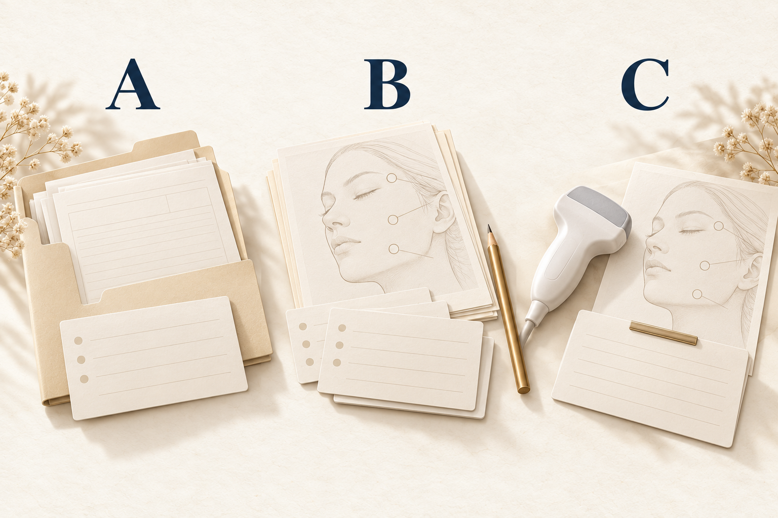
The original problem and the changes from later treatment also need to be kept apart. In areas that have been aspirated, cut or injected with medication, the way it feels and looks now may reflect more than the first filler. Keep old imaging, operation notes and pathology reports together with their dates. Then I can see the whole sequence instead of comparing your face today with an undated photo.
Photos compare the surface: how much the two sides differ, where it pulls when you smile, where the bump is. They can’t show depth or texture. You can still come in without old photos. Preparing your records simply makes the information you have more useful.
Ultrasound and pathology answer different questions
Under the right conditions, high-frequency ultrasound shows which layer something is in, how far it extends and how it relates to the surrounding tissue. Early model and clinical research described how different fillers appear on ultrasound and the local abnormalities around them. HA, Radiesse and silicone each have their own look on imaging, and together with your history that lets me estimate the general type. Recognizing certain patterns is still a long way from reconstructing every brand you’ve ever had. Ultrasound detection research https://onlinelibrary.wiley.com/doi/10.1111/j.1468-3083.2011.04047.x
The image has to be matched against your current concern. When I see what looks like a deposit, I still need to confirm it is where you feel the lump or the discomfort. When several appearances are mixed in one area, I won’t pick the part that most resembles one material and name the whole area after it. At that point, the more useful information is usually which layer it’s in, whether it is concentrated, and which vessels and nerves nearby need to be avoided.
Tissue reaction is a different question again. Granuloma is a specific pathological diagnosis. How hard something feels or a single scan can’t establish it. A retrospective biopsy study of persistent nodules showed that pathology can help tell lesions and material types apart. Those patients had already been selected for biopsy, though, so it doesn’t mean everyone with an unknown filler needs one. Pathological differential-diagnosis study https://pubmed.ncbi.nlm.nih.gov/32928687/
Taking tissue for testing is a procedure in itself. Whether it’s worth doing depends on which question it would answer and whether the answer would change treatment. If history, examination and follow-up already allow sensible management, there’s no need to run every test just to get a complete label. When the information isn’t enough, I explain what the next test is for and what its limits are.
Table 1. Different evidence answers different questions. This divides the roles of information; it does not rank test accuracy.
| Information source | What it helps establish | What it cannot establish alone |
|---|---|---|
| Original treatment and product records | The documented product, site and date | The cause of every current symptom |
| Recollections and older photographs | Sequence of events and changes in appearance or symptoms | Exact ingredients, depth or pathology |
| Ultrasound and other imaging | Location, extent and tissue relationships within the examination’s limits | Every brand, previous injection amount or complete removability |
| Tissue examination when indicated | Pathology and some material clues in the sampled area | That unsampled areas are identical, or that everyone needs biopsy |
What’s still uncertain belongs in the plan
I sort my assessment into three levels: what is confirmed, what is the more likely explanation, and what is still unresolved. Confirming a localized lesion doesn’t mean we know every component. Knowing that a certain material was injected doesn’t mean it causes every symptom you have now. Keeping these levels separate stops a reasonable guess from gradually sounding like a firm diagnosis.
There is still an order to the next steps. Something changing quickly or currently inflamed needs a decision about immediate treatment first. A stable concern about appearance leaves more time to compare options. An unconfirmed material name shouldn’t hold up the symptom assessment you need. Equally, being in a hurry to fix things shouldn’t mean skipping a question that affects safety.
If removal comes up, I first explain which area we would treat, what we plan to leave, and how we would adjust if what we find differs from what we expected. Material may be scattered through the tissue, so I can’t promise to clear all of it. At Liusmed Clinic, removal is done under ultrasound guidance through a pinhole of about 2 mm, with no incision and no stitches. If the area is large or the material is spread out, I treat it in sections and in stages. The aim is as much improvement as can be done safely, not 100 percent. FDA information on removal also notes that some fillers are difficult or impossible to remove and that the procedure itself carries risk. FDA information on removal limitations https://www.fda.gov/medical-devices/aesthetic-cosmetic-devices/dermal-fillers-soft-tissue-fillers
A trustworthy explanation doesn’t need to pretend to know everything. It should tell you what the current assessment is based on, which unknowns are most likely to change the plan, and how we intend to clear them up. Confident lines like "one scan will tell us" or "anything can be removed" skip exactly the part that matters most.
Bring what you have, without guessing the answer first
Before your consultation, put the areas and dates on one page, then attach whatever original records, photos, imaging or pathology reports you can get. Add a note on the symptom that bothers you most and the look you want to keep. This article is about reasoning when the material is unknown. For general questions about costs, follow-up visits and aftercare, use the first-visit checklist in this series.
You can keep your records and bring them to the appointment, or first ask the clinic how it receives medical information. Don’t post medical records or photos that show your face in public comments or reviews. If you have unusual pain that is getting worse quickly, skin discoloration or vision changes, seek medical care directly instead of waiting until your records are complete. If things are stable, starting from what can be confirmed will get you to a plan faster than guessing the material over and over.
Explore assessment and preparation when the filler material is unknown https://www.minimalcutsurgery.com/en/services/repair/filler
References and evidence context
[1] Murray et al. 2021. Guideline for the Safe Use of Hyaluronidase in Aesthetic Medicine, Including Modified High-dose Protocol. https://pmc.ncbi.nlm.nih.gov/articles/PMC8570661/
Professional guidance on hyaluronidase, targeting and medicine-related risk; no doses are reproduced.
[2] Wortsman et al. 2012. Ultrasound detection and identification of cosmetic fillers in the skin. https://onlinelibrary.wiley.com/doi/10.1111/j.1468-3083.2011.04047.x
Model and clinical observational research on filler appearances and associated abnormalities, not certain identification of a commercial brand.
[3] Modarressi et al. 2020. Granulomas and nongranulomatous nodules after filler injection: Different complications require different treatments. https://pubmed.ncbi.nlm.nih.gov/32928687/
Retrospective study of 26 patients selected for biopsy of persistent nodules; distinguishes a clinical nodule from granuloma, not population complication rates.
[4] U.S. FDA. Dermal Fillers (Soft Tissue Fillers). https://www.fda.gov/medical-devices/aesthetic-cosmetic-devices/dermal-fillers-soft-tissue-fillers
Official information on removal limitations and emergency warning signs. U.S. approval is not approval in other jurisdictions.
Dr. Ta-Ju Liu
