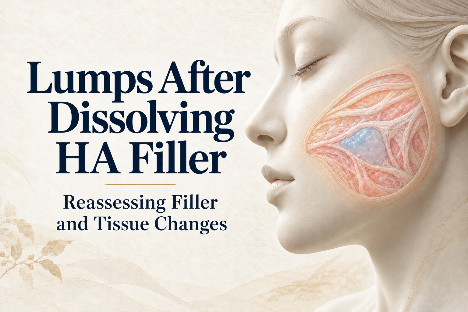
"I’ve had the filler dissolved. In the mirror it looks a bit smaller, but when I touch it, the lump is still there. Did it not all dissolve? Should I just have another round?" Quite a few people come to my clinic with two or three rounds of hyaluronidase already on their record, each one given for the same reason: let’s try a little more.
I usually ask them to pause. Being able to feel something tells us something is there. It doesn’t tell us what it is. Hyaluronidase only breaks down hyaluronic acid. If what remains is not HA, or the HA is walled off where the enzyme can’t reach it, more rounds just go in circles.
Hyaluronidase only works on hyaluronic acid
Hyaluronidase is the enzyme that breaks down hyaluronic acid (HA). Of the fillers in common use, HA is the one an enzyme can dissolve. Collagen stimulators such as Sculptra, Ellansé and Radiesse give hyaluronidase nothing to work on. If another material was used in the same spot, or what you feel is mainly a change in the surrounding tissue, repeating the injection won’t reach the real problem. Hyaluronidase safety guideline https://pmc.ncbi.nlm.nih.gov/articles/PMC8570661/
Even confirmed HA does not all dissolve equally well. A 2024 laboratory study compared 16 HA products and found they broke down at different rates. A gel in a test tube has no skin, no tissue distribution and no treatment history, though, so the results can’t be converted into how many sessions you need, what dose, or how long it will take to go down. In-vitro comparison https://pubmed.ncbi.nlm.nih.gov/38366708/
Another cause that often gets missed is simply not reaching it. Filler can shift over time. If the injection is aimed at the original injection site, or wherever the lump feels like it is, the needle can easily land beside it. That lump isn’t resistant to dissolving. The enzyme never touched it.
I also see the opposite problem in clinic. After several rounds of hyaluronidase, the lump has barely shrunk, but the tissue around it has already been thinned and dented. The enzyme can’t tell filler from the hyaluronic acid your skin naturally contains, and it breaks down whatever it spreads to.
What the remaining lump might be
People often use "granuloma" to mean any lump, but the two are not the same. A lump or nodule is something you can feel or see. A granuloma is a specific tissue change that has to be confirmed under the microscope. Biopsy studies of persistent nodules have found both granulomatous and nongranulomatous lesions. A visible bump doesn’t tell you its pathology, and a lump that survived dissolving is not proof that a thick capsule surrounds it. Study of granulomatous and nongranulomatous nodules https://pubmed.ncbi.nlm.nih.gov/32928687/
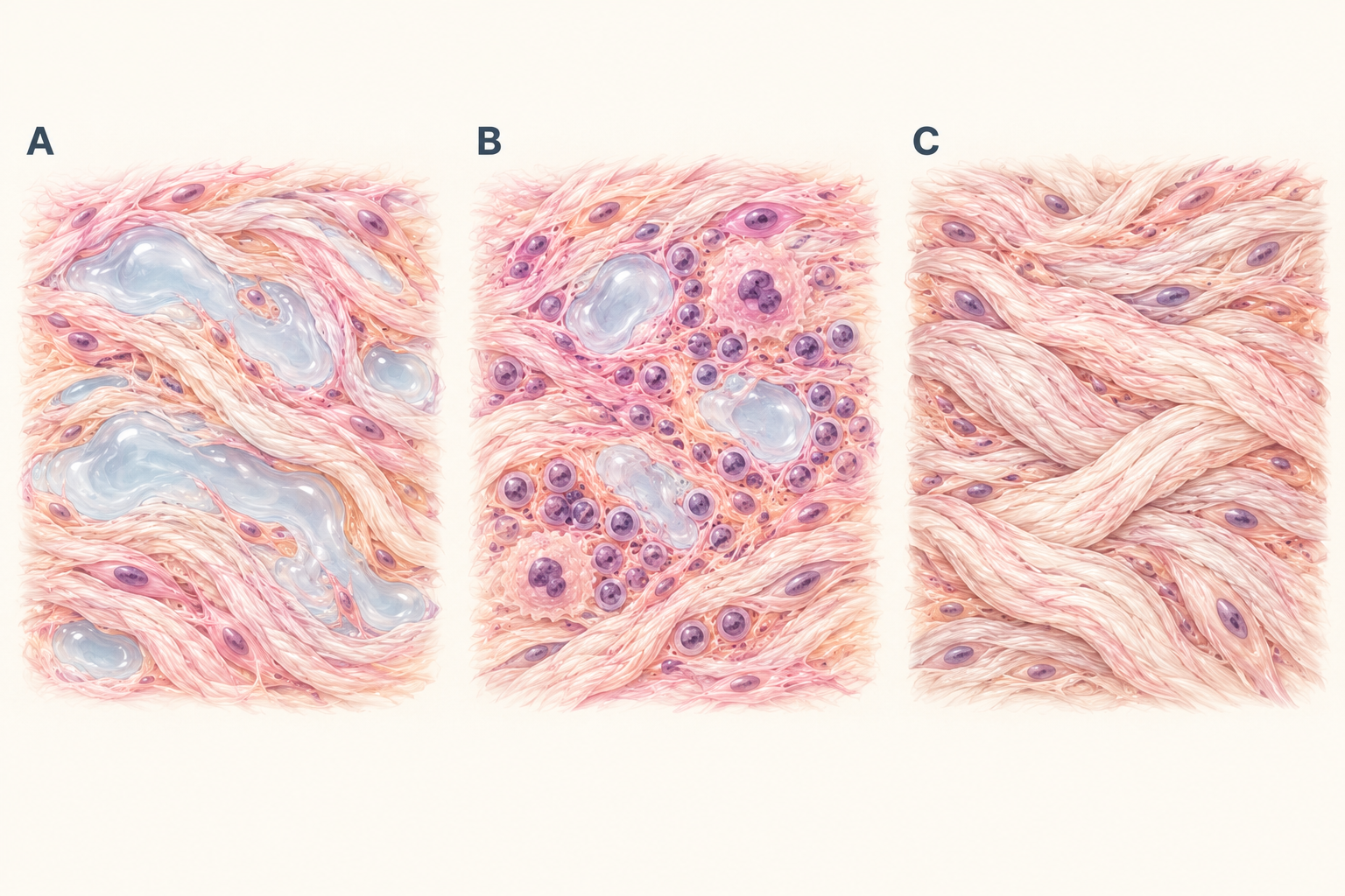
The illustration splits possible remaining material, inflammation and fibrosis into three panels to make the explanation easier. In reality all three can exist together. You won’t necessarily fit into just one panel, and material doesn’t move through them in order. What I’m looking for is the part that contributes most to your current symptoms or contour.
"There’s still a lump" can mean different things
First, compare what you feel with what you see. For some people it is mainly something they can feel, barely visible with a relaxed face. For others, a whole area looks too full but there is no distinct lump. In the first case, aiming for a result you can’t feel at all may cost far more in treatment than it gains in appearance. In the second, focusing on one small spot won’t explain the shape of the whole area.
Next, look for signs of inflammation. A bump that has stayed stable and an area that keeps turning red, warm or painful call for a different order of priorities. Consensus guidance on delayed HA reactions considers several presentations and stresses individual assessment of cause and treatment. Redness and swelling don’t automatically mean infection, and having no fever doesn’t let you rule infection out yourself. While an area is red, swollen, warm and painful, I deal with the inflammation first. That is not the time to inject or to remove. Consensus on delayed HA reactions https://pubmed.ncbi.nlm.nih.gov/32440404/
It also matters exactly what went down after dissolving. If the overall puffiness settled but left a more obvious firm spot, record the change in appearance and the change in feel separately. Even then, that doesn’t tell you what the spot is made of. If a new hollow appears after treatment, compare it with the original contour, where the injections went and when. One photo can’t tell you which tissue was affected.
The table below is meant to help you describe "it didn’t go away" more precisely. When someone tells me that, I need to know which part didn’t improve.
Table 1. Identify what has not improved. These are observations and assessment questions, not diagnoses or treatment instructions.
| What you notice | Why describing it this way helps | What it doesn’t tell us yet |
|---|---|---|
| Swelling has gone down but a localized firm spot remains | Tracks overall appearance separately from the local feel | Whether the whole spot is residual HA |
| A fixed bump with no long-term change in redness, warmth or pain | Describes how stable it is and how far you want the appearance to improve | What material it is, judged only on stability or lack of pain |
| Repeated redness or swelling, or new pain | Flags a possible ongoing tissue reaction that needs reassessment | A self-diagnosis of infection, allergy or granuloma |
| Still uneven, but no single lump can be found | Means looking at the whole area’s distribution and how the contours blend | Whether removing one lump would fix the overall appearance |
Before the next injection, look closely at the last one
"I’ve had hyaluronidase several times" doesn’t tell me much. The date of each session, the exact site, the medicine used and the dose written in the records show which problem was being treated at the time. Put the original filler, the first sign of trouble, the later dissolving and any other injections or surgery on one timeline. That keeps changes from different stages from being merged into a single conclusion.
Photos are most useful for comparing contour and skin changes. Take them in similar lighting, angles and expressions, and keep the dates. If symptoms come and go, note where and how it feels during a flare. You don’t need to keep pressing hard to prove the lump is still there. Depth, texture and how it moves with expression don’t show in photos and still need an examination.
If two rounds have done nothing, I’d scan before deciding on a third. Ultrasound tells me whether what you feel matches what I see on the image, which layer it sits in, and whether it is concentrated in one spot or spread out. Research has described how different fillers look on ultrasound, including mixed appearances. So ultrasound helps locate the problem and estimate the material, but one scan can’t confirm the brand, every component or how much was injected each time. Ultrasound-pattern study https://pmc.ncbi.nlm.nih.gov/articles/PMC8252486/
If no one can say what was originally injected, hold off on the next injection. Assuming it is all HA and counting every disappointing result as a "dissolving failure" easily sends things further off course. I go back to the original records and separate what is documented from what is only the most likely explanation right now.
The next step should target what is actually left
If ultrasound shows HA that hasn’t fully dissolved, and the last round made it a little smaller, another injection aimed precisely at the residue makes sense. If two or more rounds produced no change at all, or I can see a capsule around it, I usually recommend stopping and assessing for removal instead. If inflammation or a different lesion is the main issue, that gets treated first. A stable finding with no reason to act now can be followed with a clear observation plan. Being unhappy with the last round doesn’t mean the next one should be stronger or bigger.
Follow-up needs content too. Are we tracking size, appearance, symptoms or a test result? Which changes would make us change the plan? Agreeing on this up front separates waiting for tissue to settle from putting off a question that hasn’t been answered. "Let’s wait a bit" on its own leaves patients unsure what they’re waiting for.
If removal comes up in the end, the conversation turns to how much to treat and which tissue to keep. Making a bump smaller and clearing out all the material are two different goals. After removal, an area that was being propped up may look more hollow, and how it blends with its surroundings matters too, so the amount removed is not a measure of how good the repair is. At Liusmed Clinic, removal is done under ultrasound guidance through a pinhole of about 2 mm, with no incision and no stitches. The first session usually removes about 30 to 50 percent. For most fillers, two to three sessions remove about 80 to 90 percent, with about 3 months between sessions. It is not 100 percent, and it varies from person to person. I also don’t aim to take everything out. I follow your natural lines, take only the part that sticks out, and smooth what remains. The FDA also cautions that reducing or removing filler carries its own risks and that some materials are hard to remove completely. FDA information on fillers https://www.fda.gov/medical-devices/aesthetic-cosmetic-devices/dermal-fillers-soft-tissue-fillers
Before your next visit, write one sentence about the symptom that bothers you most and one about the change you most want in your appearance. That is far more specific than "dissolve all of it." Then ask the doctor to spell out the most confident assessment, what evidence is still missing, which options exist, and what new problems each option could bring. Once that is clear, you can look back later and check whether the plan is on track.
New warning signs should not wait for a routine schedule
Unusual or steadily worsening pain, pale or gray-blue skin, or changes in vision need immediate medical attention. Increasing redness, warmth, pain, discharge or fever also need prompt assessment. Do not inject more hyaluronidase, puncture the area or massage it hard yourself, and do not delay care while waiting for a routine appointment or a reply to photos. This article explains how to think it through. It cannot replace an examination of your current symptoms.
Explore assessment for persistent concerns after HA filler dissolving https://www.fillerrevision.com/en/services/repair/filler
References and evidence context
[1] Murray et al. 2021. Guideline for the Safe Use of Hyaluronidase in Aesthetic Medicine, Including Modified High-dose Protocol. https://pmc.ncbi.nlm.nih.gov/articles/PMC8570661/
Professional guidance on hyaluronidase, targeting and medicine-related risk; no doses are reproduced.
[2] Faivre et al. 2024. Comparison of Hyaluronidase-Mediated Degradation Kinetics of Commercially Available Hyaluronic Acid Fillers In Vitro. https://pubmed.ncbi.nlm.nih.gov/38366708/
In-vitro comparison of 16 HA products; supports differences in degradation, not human dose, session count or success rate.
[3] Modarressi et al. 2020. Granulomas and nongranulomatous nodules after filler injection: Different complications require different treatments. https://pubmed.ncbi.nlm.nih.gov/32928687/
Retrospective study of 26 patients selected for biopsy of persistent nodules; distinguishes a clinical nodule from granuloma, not population complication rates.
[4] Philipp-Dormston et al. 2020. Global Approaches to the Prevention and Management of Delayed-onset Adverse Reactions with Hyaluronic Acid-based Fillers. https://pubmed.ncbi.nlm.nih.gov/32440404/
Expert consensus on delayed HA reactions; individualized assessment, not a protocol for every biostimulator.
[5] Urdiales-Gálvez et al. 2021. Ultrasound patterns of different dermal filler materials used in aesthetics. https://pmc.ncbi.nlm.nih.gov/articles/PMC8252486/
Original observational study of ultrasound patterns and mixed appearances; not guaranteed brand identification or complete removal.
[6] U.S. FDA. Dermal Fillers (Soft Tissue Fillers). https://www.fda.gov/medical-devices/aesthetic-cosmetic-devices/dermal-fillers-soft-tissue-fillers
Official information on removal limitations and emergency warning signs. U.S. approval is not approval in other jurisdictions.
Dr. Ta-Ju Liu
