Understand the tissue first, then decide how to repair it
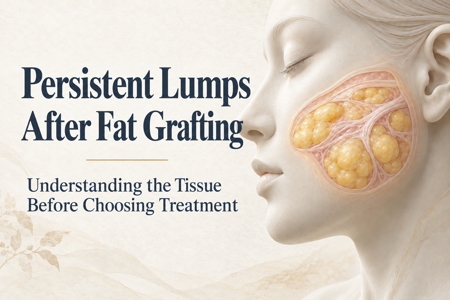
"It’s been years since my fat grafting and this lump is still here. Did the fat not absorb? Can’t you just take it out?" That is often the first thing people ask me when they come in with a lump after fat grafting. Once I examine and scan the area, though, "uneven" turns out to mean different things. For some it is a whole area that is too full. For others it is a single, separate lump under the skin. For others it barely shows until they smile and a line pulls across. What sits inside is different in each case, and so is the repair.
So I don’t stop at how hard it feels or how long ago the grafting was. What I want to know is whether there is still living fat in that area, whether there is a pocket of oil in the middle, and whether the tissue around it is inflamed or already fibrotic. Those answers decide whether we can watch it first, which part needs treatment, and what will hold the contour up afterward.
Fat from the same session can end up in different states
Your fat is your own, but after transfer it still has to grow a new blood supply. Harvested fat does not move with its original vessels attached. It has to survive in its new location on its own. Experimental work shows that within the same graft, some cells survive and some do not.[1,2] So knowing that an area was grafted does not tell you that all of it is still alive, or that all of it has died.
When fat cells die, they release oil. Some is cleared by the body and some collects. Once tissue walls it off, it becomes an oil cyst. An inflammatory reaction to that oil is a lipogranuloma (fat granuloma). More fibrous tissue is fibrosis. Calcium deposits are calcification.[2–4] People often use these words interchangeably. To me they are different findings, and they point to different treatment.
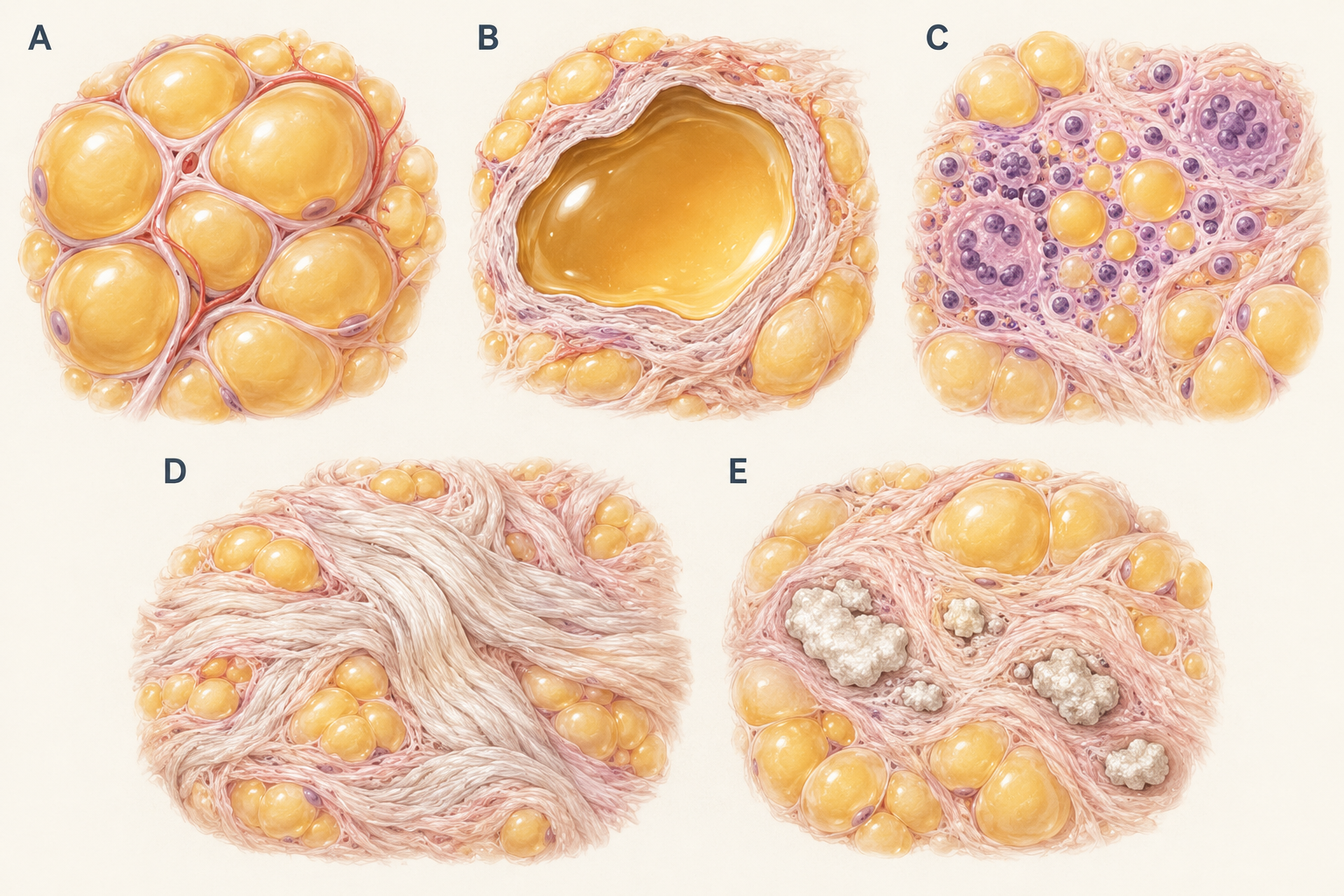
In the illustration, compare B, C and D. An oil cyst describes what has collected inside. A lipogranuloma describes the reaction around it. Fibrosis makes the tissue feel tighter and firmer. One lump often contains more than one of these, and there may be normal fat right beside it that should stay. Draining some oil does not necessarily fix the appearance or the symptoms.
| Tissue finding | What it means | What I look at when assessing it |
|---|---|---|
| A Living fat | Fat that is still alive. When the amount or position is wrong, it simply looks too full. | Whether this is a broad volume problem, and whether the area will blend with its surroundings after reduction. |
| B Oil cyst | A collection of oil, sometimes surrounded by a fibrous wall. | The oil, the wall and the reaction around it. Draining the oil does not necessarily clear the surrounding changes. |
| C Lipogranuloma | A granulomatous inflammatory reaction to oil. | The inflammation and symptoms first, then whether to observe, use medication or treat the lesion. |
| D Fibrosis | More fibrous tissue, which feels tight and firm. | Its size, and how firmly it is stuck to nearby tissue. It is not as simple as draining a pocket of oil. |
| E Calcification | Calcium deposits, often alongside other changes. | Confirmation on imaging. Feeling hard does not mean calcium, and firmness should not decide how much to treat. |
Table 1. A guide to tissue differences, not a self-diagnosis tool. Components may coexist; treatment decisions depend on examination and any indicated investigations.
"It feels like a stone. Does that mean it has calcified?" Firmness is only a clue, because fibrosis can also make tissue hard. A study of oil cysts after breast fat grafting did find chronic inflammation, fibrosis and calcium together. That was pathology from three patients, though. It shows these changes can overlap, not that every facial lump follows the same course.[3]
If it has lasted for years, map out how it changed
"I’ve always been able to feel it, but it hasn’t changed in years" and "It was stable, then recently got bigger and started to hurt" are two different histories to me. Before your visit, write down the date of grafting, where the fat was placed, and when you first felt the lump, as separate items. If you had other fillers, aspiration, injections or surgery in between, put them on the same timeline. I need to tell whether what I see now comes from the original grafting or from later treatments layered on top.
Location matters too. A small lump under the eyelid, a deeper one in the cheek and a bulge you can see on the surface carry different difficulty and risk, even when they are about the same size. Photos record appearance but not depth or texture. A lump you can feel but others barely notice is almost impossible to show in a single front-on photo.
Ultrasound is my first step in assessing a fat-graft lump, and I go in with specific questions. Which layer is it in? Is it oil, fibrous tissue or calcium? Which nerves and vessels are nearby? Grafted fat grows into the surrounding tissue, and the border is often unclear, which is why I don’t make the call by touch alone. Imaging gives clues about position and composition. Seeing an outline still does not tell you the full pathology. If it remains unclear, whether to take a tissue sample is a separate decision.[4,5]
Around the eye, eye function also comes into it. A lump that comes with eyelid swelling or difficulty opening the eye takes priority over a small spot you can merely feel. When someone has a bulge in the upper eye socket after fat grafting, I am deliberately conservative the first time and reassess after full recovery before deciding on a second session, because removing too much here turns a bulge into a hollow. If the assessment shows a stable lump with no reason to act now, observation is a legitimate option. We should agree on which changes mean you come back sooner, rather than leaving you with "let’s just wait and see."
Decide first what should stay
Before we talk about methods, I ask which one thing you most want to change: pain, swelling that keeps coming back, a lump you can feel, or a bulge you can see. These often appear together, but one treatment may not solve all of them. Once a lump gets smaller, the area it was propping up can look hollow. A smoother surface does not mean the painful lump has been dealt with.
If inflammation is the main problem, removal is not always the first move. Park and Kim reviewed 27 cases of periorbital lipogranuloma. Some resolved during observation, some improved after local steroid injection, and some recurred after treatment. This was a small, nonrandomized retrospective study, with tissue confirmation in only three cases, so it is not a general rule for oil cysts, calcium or excess living fat.[4] Calcification is a different matter. Once calcium has formed, it usually doesn’t break down on its own, and simply waiting rarely changes how hard or how large it is.
When treatment is needed, the method depends on what is inside. A lump that is mostly oil is a different job from one that is mostly dense fibrous tissue or calcium. How well defined it is, and how tightly it is stuck to the tissue around it, also change the approach. At Liusmed Clinic, I remove fat-graft lumps and localized overfilling under ultrasound guidance through a pinhole of about 2 mm, with no incision and no stitches. When the area is large or spread out, I treat it in sections and in stages, still through pinholes. Oil cysts, fibrotic masses and calcified nodules each look different on ultrasound, and I adjust the removal to match.
A lump in the face is not sitting in an empty pouch. Next to it may be normal fat that supports your contour, or nerves, blood vessels and the parotid gland. A 2026 case report described a salivary collection (sialocele) after a deep cheek fat-graft lesion was excised. One case cannot tell you how often this happens. It is enough to show that even open surgery, where the lesion is in direct view, cannot promise to remove everything without affecting what is next to it. Whether nearby structures are protected belongs in the conversation alongside how much can be removed.[5]
That is what I care about most when removing fat-graft lumps. I do liposuction and fat grafting, and I also repair liposuction that went wrong. Removing a fat-graft lump is a bit like precise fat grafting in reverse. I map the raised area first, account for how it pulls when you change expression, and reassess once the swelling settles, so that I take neither too much nor too little. The goal is to follow your natural lines, take only the part that sticks out, and smooth what remains. I don’t chase getting every last bit out. Some people come to me after removal elsewhere left them lumpy. Having had something removed is not the same as having it removed evenly.
So at your consultation, besides asking how much can be removed, ask what will be left and why. If I suggest leaving some, treating in stages or watching first, I will explain what that protects and which results will guide the next step. The reason for staging is practical: removing too much in one go easily leaves hollows or unevenness that are harder to fix than the original lump. Every repair carries trade-offs too, including residual material, recurrence, changes in sensation, infection, and new irregularity or asymmetry, and these should be discussed before anything is done. The pinhole is small, but this is still removal, so swelling and bruising will happen, and I won’t tell you the recovery is short. Swelling and bruising usually settle in about 2 to 3 weeks. The pinholes heal in about 2 weeks and fade over about 2 months. The final result takes about 3 months. Everyone heals differently.[5,6]
Too much living fat is a separate issue. It is a matter of volume and distribution. A treatment that calmed an inflamed nodule has not been shown to reduce excess living fat precisely. When reducing it, I look at how the area blends with its neighbors and how it moves with expression. What we need to agree on is which part of the contour to refine and how much fullness you can accept leaving, including the chance of further treatment, rather than setting a fixed percentage to remove.
What to bring to your next consultation
Bring dated photos, any treatment records you can get, and previous imaging or pathology reports. Write one sentence about the appearance that bothers you most and one about your actual symptoms. Then ask the doctor what this is most likely to be, what evidence is still missing, what happens if you leave it alone, and what new problems treatment could bring. Deciding to aspirate, inject or cut before the tissue is understood gets the order backwards.
New or worsening redness, warmth, pain, drainage or fever means contacting a healthcare professional promptly, without waiting for a routine cosmetic consultation. Do not puncture or squeeze the area yourself, and do not use someone else’s medication.[7]
If a lump or localized fullness after fat grafting is bothering you, you can first read how a fat-graft revision assessment works, then bring the records you have to discuss the next step.
Explore fat-graft revision assessment https://www.fillerrevision.com/en/services/repair/fat-graft
References and the limits of the evidence
[1] Eto H, et al. The fate of adipocytes after nonvascularized fat grafting: evidence of early death and replacement of adipocytes. https://pubmed.ncbi.nlm.nih.gov/22261562/ Plastic and Reconstructive Surgery. 2012;129:1081–1092. Human adipose-tissue experiments and a mouse graft model inform the biology of survival. They do not establish clinical outcomes of facial revision.
[2] Kato H, et al. Degeneration, regeneration, and cicatrization after fat grafting: dynamic total tissue remodeling during the first 3 months. https://pubmed.ncbi.nlm.nih.gov/24572875/ Plastic and Reconstructive Surgery. 2014;133:303e–313e. Mouse histology demonstrates different tissue outcomes. Its time points are not a recovery timetable for a human facial lump.
[3] Mineda K, et al. Chronic inflammation and progressive calcification as a result of fat necrosis: the worst outcome in fat grafting. https://pubmed.ncbi.nlm.nih.gov/24776542/ Plastic and Reconstructive Surgery. 2014;133:1064–1072. Pathology and imaging of oil cysts from three breast fat-grafting patients show coexisting inflammation, fibrosis and calcium. This does not predict every facial lesion.
[4] Park JY, Kim N. Periorbital Lipogranuloma after Facial Autologous Fat Injection and Its Treatment Outcomes. https://pmc.ncbi.nlm.nih.gov/articles/PMC4742640/ Korean Journal of Ophthalmology. 2016;30:10–16. A retrospective series of 27 periorbital cases, with histology in only three. Nonrandom treatment selection prevents a fair comparison of treatment success.
[5] Ma IZ, et al. Sialocele following excision of delayed-onset inflamed facial fat graft: A case report. https://pubmed.ncbi.nlm.nih.gov/41824886/ Medicine. 2026;105:e48037. A single facial case highlights nearby-structure risks during deep revision. It shows a possible complication, not its frequency or any clinic’s surgical results.
[6] American Society of Plastic Surgeons. Liposuction Risks and Safety. https://www.plasticsurgery.org/cosmetic-procedures/liposuction/safety Patient information. Professional-society information for general liposuction risk discussions, not a comparative trial of facial fat-graft revision.
[7] Centers for Disease Control and Prevention. Surgical Site Infection Basics. https://www.cdc.gov/surgical-site-infections/about/index.html Patient information. Official postoperative safety information supporting prompt assessment of redness, pain, drainage or fever.
Article information
English search title Lumps after facial fat grafting: oil cysts, granulomas and calcification
English description A lump that lasts years after facial fat grafting may be living fat, an oil cyst, a fat granuloma, fibrosis or calcium. Ultrasound shows which one, and that decides whether to watch, investigate or treat, and what normal tissue and contour should stay.
Dr. Ta-Ju Liu
