
"I can feel it, but my doctor said there’s nothing there." I hear this often in clinic. Other people see a dark area on the screen and ask right away: "Is that what I had injected? Now that you’ve found it, can you get it all out?"
I usually answer with a question of my own: over all these years, has any doctor scanned your face first? Between "there’s a dark patch" and "it can all come out" sit several decisions. Does the spot on the screen match the lump you can feel? What is probably inside it? Which tissue around it needs to stay? You have to see it before you can treat it safely. Without imaging, I won’t tell anyone "yours can be removed" or "yours can’t."
Start by matching your concern to the image
A bulging cheek can mean a distinct lump under the skin, a whole area that feels too full, or unevenness that only shows when you smile. So before I scan, I ask you to point it out: where it is, when it started and whether it has been growing or shrinking. If you’ve had several treatments, I need to know where each one went. Finding one deposit and blaming it for every change in your face leads the assessment in the wrong direction.
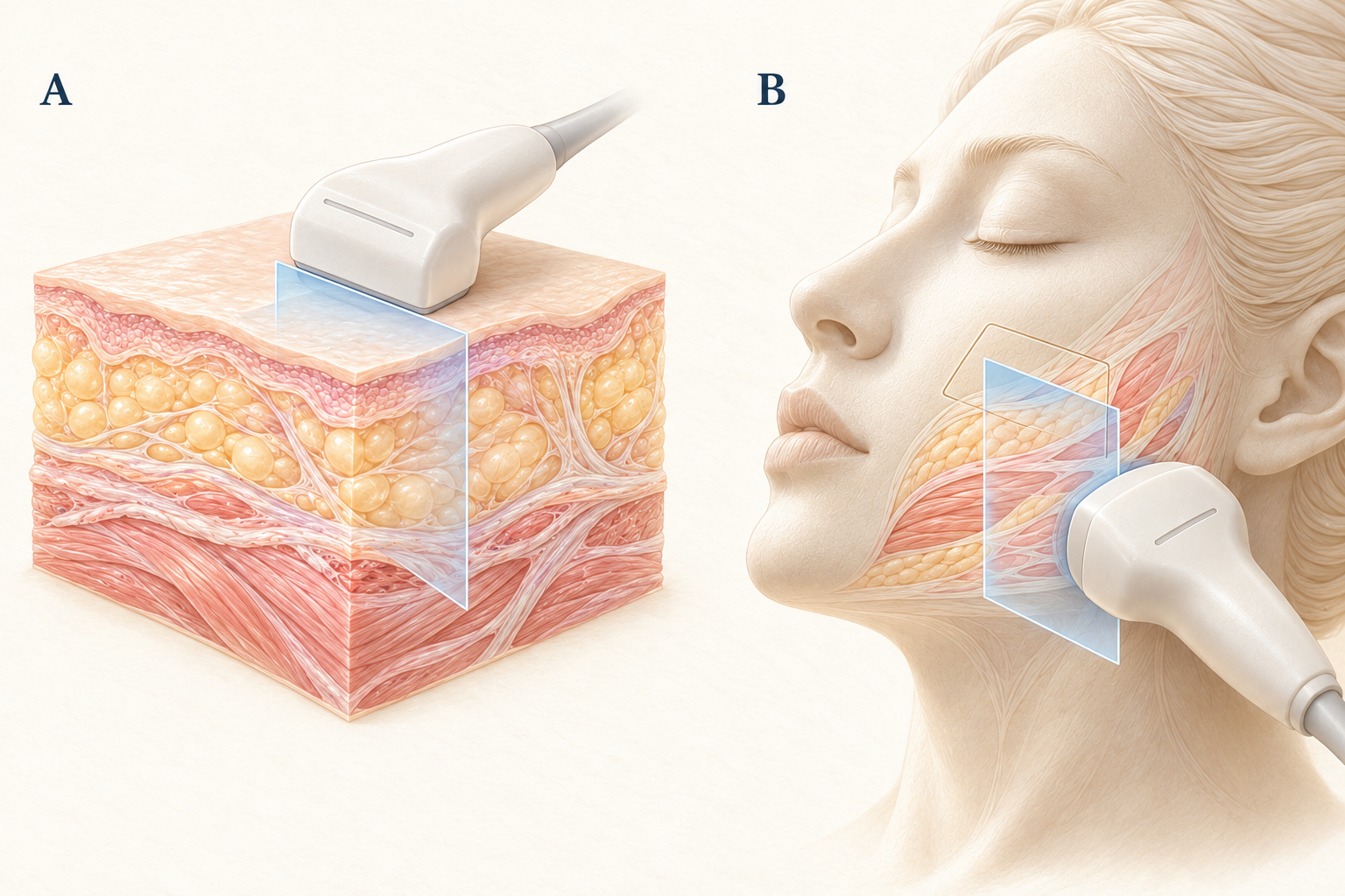
I also don’t scan only the spot you point to. Filler migrates, so material injected in one place can turn up next to it. Quite a few people find out after a scan that there is more in their face than they remembered, and not where they thought. Ultrasound shows one cross-section at a time. The probe has to move, and images from different directions have to be matched to the surface. A single screenshot is not a map of the whole face. When needed, Doppler adds information about blood flow. Whether to add it, and how to read it, depends on the question the scan is meant to answer. How ultrasound and Doppler work https://www.radiologyinfo.org/en/info/genus
For a lump you can feel, I’m looking at which layer it sits in, where its edges are and how close it is to nearby vessels and nerves. For broader unevenness, the image has to be read alongside the overall contour, the difference between sides and how the face moves. Imaging fills in what I can’t see from the outside. Which part of the contour is most worth improving still comes back to your face and what bothers you.
Wortsman and colleagues combined a porcine skin model with clinical observations to describe filler deposits visible on ultrasound and abnormalities in the surrounding tissue. It shows that ultrasound can see the material. It does not show that every visible deposit needs treatment. Your doctor should be able to explain how strongly a finding relates to your symptoms. Original study https://onlinelibrary.wiley.com/doi/10.1111/j.1468-3083.2011.04047.x
An image gives clues about the material, not a product label
Some materials are easier to recognize on ultrasound. But which brand, which category of material and what the tissue looks like now are three different questions. Recognizing a likely category still doesn’t tell me which product was used years ago, how much was injected or how each session was done.
In a retrospective observational study of 60 people, 12 showed mixed patterns, and the appearance of hyaluronic acid (HA) was described as changing as it integrated with tissue. After several treatments, or after time has passed, the picture is rarely as simple as it was right after injection. Experienced operators performed that study. Its pattern classification is not a brand test that any machine or any operator can reproduce. Study of filler ultrasound patterns https://pmc.ncbi.nlm.nih.gov/articles/PMC8252486/
So "consistent with a certain material" on a report and "confirmed as a specific product by the original records" carry different levels of certainty and should be written separately. When the history and the images don’t agree, saying clearly what remains uncertain is more useful than forcing a name onto the finding. That uncertainty also makes treatment more cautious: looking like one category doesn’t justify a method that only suits that category.
The reverse holds too. Not seeing a distinct collection doesn’t mean "nothing is left." Which areas were scanned, which layers were seen clearly and whether something else could explain the symptoms all need to be stated. A scan that finds nothing is still an answer, as long as you know which question it answered.
What I want is information that changes a decision
I check whether a finding is useful with one question: if it’s true, what changes in the plan? Clearer localization means I can plan where to treat. A finding that doesn’t match the symptoms sends me back to look for another cause. If there’s no reason to treat yet, a baseline image is kept for comparison later.
Assessment and guidance are also different things. Assessment gathers information. Guidance uses real-time imaging during a procedure to confirm where the target and the instrument are. Having an ultrasound assessment doesn’t mean every later step happens under imaging, and having guidance doesn’t make a procedure suitable for every material. Ultrasound can guide aspiration or sampling in general practice; whether a particular filler revision needs that is a separate clinical decision. In my practice, extraction is done under real-time ultrasound guidance through a pinhole of about 2 mm, with no incision and no stitches, and I perform every extraction myself. Uses of real-time imaging https://www.radiologyinfo.org/en/info/genus
The question people skip most often is where treatment should stop. The suspicious area on an image isn’t necessarily the area that can be removed safely. Normal fat, skin and other soft tissue also hold up the contour. The more that comes out, the higher the cost in hollowing, asymmetry and injury to nearby structures. The FDA also notes that some fillers are difficult to remove and that removal may need additional procedures with their own risks. Limitations of filler removal https://www.fda.gov/consumers/consumer-updates/dermal-filler-dos-and-donts-wrinkles-lips-and-more
That’s why, before an extraction, I map the whole face, mark the raised areas and take into account how expression pulls on them. What I’m aiming for is a balanced overall contour. An overfilled area doesn’t need to be emptied; I remove the parts that stand out and smooth what stays. Having had something removed isn’t the same as having it removed thoroughly, and even less the same as having it left smooth. If, after the scan, I suggest treating only part of an area, staging the treatment or watching for now, please don’t judge that only by whether everything came out. The first session usually removes about 30 to 50 percent. With most fillers, two to three sessions can remove about 80 to 90 percent. It is not 100 percent, and it varies from person to person. Sessions are usually about three months apart.
Table 1. Linking questions about the image to clinical decisions. This is a discussion aid, not a guide to reading your own scan.
| Question to clarify | Information ultrasound may add | Decision still required |
|---|---|---|
| Where is the lump I can feel? | Identifiable tissue layer, extent and neighboring structures | Whether it explains the symptoms and needs treatment |
| Which material category is possible? | A pattern consistent with certain materials, or a mixed pattern | Whether original records confirm it; brand or injected volume cannot be guaranteed |
| Could a targeted procedure help? | Target location and, where appropriate, real-time guidance | The indication, material differences and the risks of the procedure itself |
| How much could be treated? | Visible lesion and its relationship to surrounding tissue | Tissue preservation, residual material and contour trade-offs |
| How can changes be compared later? | Baseline images with location notes | Purpose of follow-up, symptom changes and reasons for reassessment |
What can’t be seen clearly belongs in the conclusion
Ultrasound has physical limits. Sound travels differently through different tissues, and structures like bone block the view. The machine, the probe, the scanning direction and the reader’s experience all affect how much is seen. One thing I want to say up front: owning a machine and knowing how to read it are two different things, and the difference is whether the operator understands the anatomy of the face, layer by layer. You don’t need to know the settings, but you can ask three things: who did the scan, who interpreted it and which areas couldn’t be seen clearly this time. General ultrasound limitations https://www.radiologyinfo.org/en/info/genus
Imaging shows shape and location, but it isn’t a tissue biopsy or a culture. With recurring redness, swelling or pain, or when the history and the images just don’t fit, the missing piece of the answer should decide whether other imaging, lab tests or sampling are needed. Another test should fill a gap that changes treatment, not simply add to the list.
Emergency signs come first. Sudden changes in vision, unusual or worsening pain, or skin that turns unexpectedly pale, gray or blue need immediate medical care, not a routine scan appointment. An earlier image that looked fine cannot rule out symptoms that appear later. FDA warning signs after injections https://www.fda.gov/medical-devices/aesthetic-cosmetic-devices/dermal-fillers-soft-tissue-fillers
Leave with a result you can build on
After the scan, the things worth keeping are the date, the side and area, the main findings, the limits of interpretation and the recommendation for what comes next. If you’ll want to compare later, images labeled with location and orientation, plus a formal report, are far more useful than an unlabeled black-and-white screenshot on your phone.
A useful conclusion can include uncertainty, but it shouldn’t stop at "something is there" or "nothing is there." Afterward you should know which finding best explains your concern, which questions remain open and whether the next step is monitoring, more assessment or considering treatment.
If you’re comparing filler revision options, start with how the assessment works, then bring your past records and images so we can go through which information will actually change your treatment choices. https://www.minimalcutsurgery.com/en/services/repair/filler
References and evidence context
[1] Wortsman et al. 2012. Ultrasound detection and identification of cosmetic fillers in the skin. https://onlinelibrary.wiley.com/doi/10.1111/j.1468-3083.2011.04047.x
Porcine model and clinical observations supporting visualization of filler deposits and associated abnormalities; not evidence of removal effectiveness or of a need to treat every deposit.
[2] Urdiales-Gálvez et al. 2021. Ultrasound patterns of different dermal filler materials used in aesthetics. https://pmc.ncbi.nlm.nih.gov/articles/PMC8252486/
Retrospective observational study of 60 people supporting mixed patterns and changing HA appearances; not a brand-identification accuracy estimate, a universal operator result or a removal rate.
[3] ACR and RSNA. RadiologyInfo. General Ultrasound. https://www.radiologyinfo.org/en/info/genus
Professional-society patient information on cross-sections, Doppler, real-time guidance and general physical limits; not a trial of facial revision outcomes.
[4] U.S. FDA. Dermal Fillers Soft Tissue Fillers. https://www.fda.gov/medical-devices/aesthetic-cosmetic-devices/dermal-fillers-soft-tissue-fillers
Official patient safety information on removal limitations, risks and urgent warning signs. U.S. approval status is not generalized to other jurisdictions.
[5] U.S. FDA. Dermal Filler Dos and Don’ts for Wrinkles Lips and More. https://www.fda.gov/consumers/consumer-updates/dermal-filler-dos-and-donts-wrinkles-lips-and-more
Official consumer information on the difficulty of removing some materials, possible additional procedures and the risks of removal.
Dr. Ta-Ju Liu
